
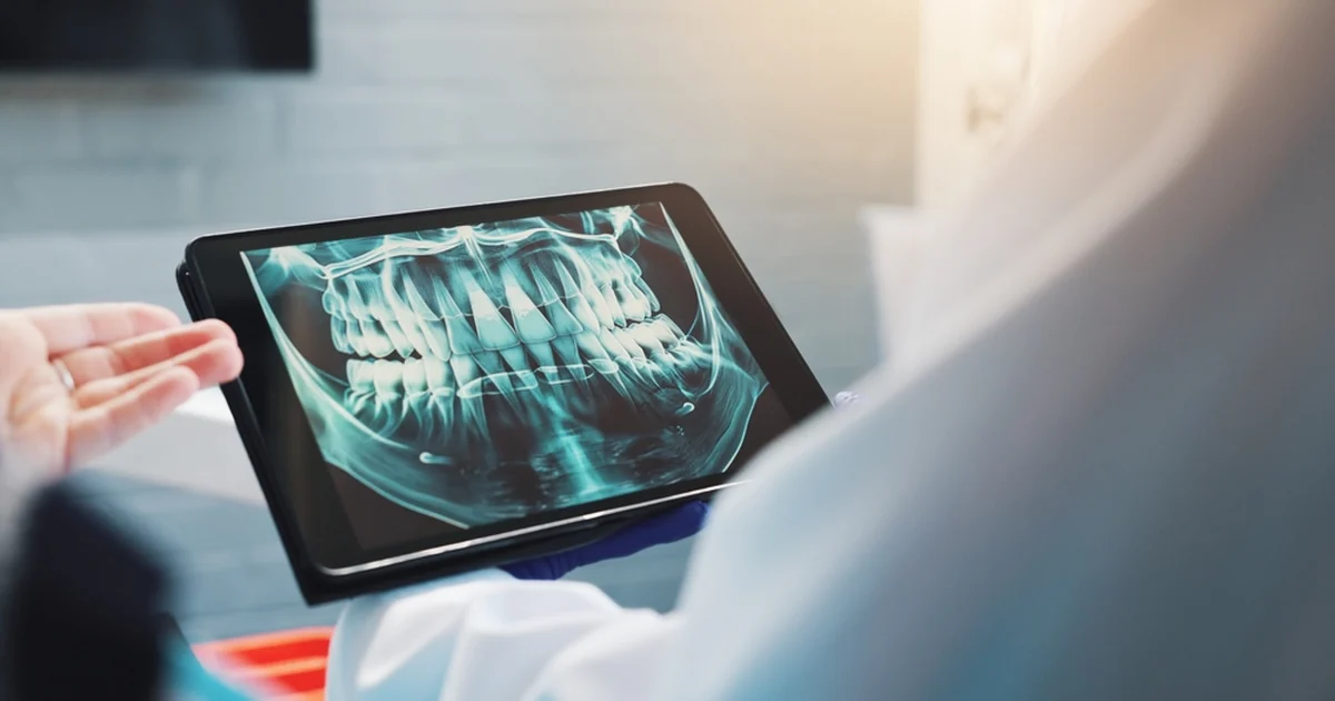
An examination tells us a great deal, but there are limits to what any dentist can see by looking. Roughly a third of each tooth sits beneath the gum, the surfaces between teeth are hidden from view, and the bone supporting your teeth is invisible entirely. X-rays fill those gaps.
They are also a common source of questions, particularly about radiation. Here is a straightforward explanation of what we use, why, and how often.
What X-Rays Reveal
- Decay between teeth, which cannot be seen directly and is one of the most common findings
- Decay beneath existing fillings and crowns
- Bone levels around each tooth, which is how we assess the extent of gum and periodontal disease
- Infections at the tip of a root, often before any symptoms appear
- Cysts and other changes in the jaw
- Position of unerupted teeth, particularly wisdom teeth
- Root shape and canals, which guides root canal therapy
- Bone volume and anatomy for planning dental implants
- Development of adult teeth in children
Many of these findings are silent. Finding a cavity between two teeth on an X-ray means a small white filling; finding the same cavity when it starts to ache often means a crown or root canal therapy.
The Types We Use
Bitewings
Small images showing the crowns of the upper and lower back teeth together. These are the standard images for detecting decay between teeth and checking bone levels.
Periapical
A single-tooth image showing the whole tooth including the root tip and surrounding bone. Used when investigating pain, infection or a specific tooth.
OPG
A digital OPG is a panoramic image showing all the teeth, both jaws, the jaw joints and surrounding structures in one picture. It is used for assessing wisdom teeth, planning implants, reviewing overall development and investigating jaw concerns. It is not detailed enough to replace bitewings for detecting small cavities.
3D Imaging
Cone beam CT produces a three-dimensional image and is used selectively, most often for complex implant planning or assessing the relationship between a wisdom tooth and the nerve. It involves a higher dose than the images above, so it is reserved for cases where the extra information genuinely changes the treatment.
How Often Are X-Rays Needed?
There is no fixed schedule, and X-rays should not be taken automatically at every visit. The interval is based on your individual risk, your age and the current state of your teeth and gums. New patients usually need a baseline set unless recent images are available from a previous practice.
Someone with no recent decay, healthy gums and good home care may need bitewings only every two to three years. Someone with active decay, a dry mouth, many existing restorations, periodontal disease, frequent sugary drinks or a smoking habit may need them more frequently. Children with high decay risk may need them more often than adults with low risk.
We will always explain why an image is recommended, and you are welcome to ask.
About Radiation
Modern dental X-rays use digital sensors, which need considerably less radiation than the film systems they replaced. The doses involved are small, and we use the principle of keeping exposure as low as reasonably achievable: shielding where appropriate, the smallest field of view that answers the question, and no image taken without a clinical reason.
For context, we are all exposed to background radiation continuously from the environment, and a set of dental bitewings represents a very small addition to that, broadly comparable to what you would receive on a short flight. That does not make X-rays trivial, which is why they are prescribed individually rather than routinely.
If You Are Pregnant
Tell us. Routine imaging is usually deferred during pregnancy, although X-rays can be taken safely with appropriate shielding if there is an urgent clinical need, such as investigating infection. Dental treatment itself is not something to postpone during pregnancy, as gum health often needs more attention during this time.
Digital Advantages Beyond Dose
Digital images appear on screen within seconds, can be magnified and adjusted, and are stored so they can be compared with previous images. That comparison is often the most valuable part, because a lesion that has not changed in three years is a different clinical picture from one that has grown.
They are also easy to show you, which makes a recommendation much easier to understand than a description alone.
If You Have Recent X-Rays From Another Practice
Bring them, or ask your previous practice to send them. There is no reason to repeat imaging unnecessarily, and older images give us valuable history.
Why We Explain the Reason Each Time
Imaging should always be justified by a clinical question. Before recommending an X-ray, the question we are answering is a specific one: is there decay between these teeth, has this bone level changed, where does this root sit in relation to the nerve.
If the answer would not change what we do, the image is not taken. That principle keeps exposure low without withholding information that genuinely affects your treatment.
You are entitled to ask what the image is for, what we are looking for and what happens if it is not taken. Those are reasonable questions, and the answers should be specific.
Book Your Check-Up
If you are due for a check-up and clean, we will assess whether imaging is needed and explain our reasoning. Call Precision Dental on (07) 3852 1160 or book online.